In December of 2021, the Foundation made a $1 million award to Drs. Maksim Plikus and Kyriacos Athanasiou at the University of California, Irvine to characterize an understudied cell type called the lipochondrocyte (LC), which secretes a small amount of extracellular matrix but contains a prominent intracellular lipid-filled vacuole, much like a fat cell. The investigators hypothesized that lipogenesis is critical for the structure-function relationship of LCs and that, analogous to “bubble wrap,” a tissue can acquire cartilage-like biomechanics via tightly packed, lipid-filled cells instead of voluminous, “packing foam”-like extracellular matrix.
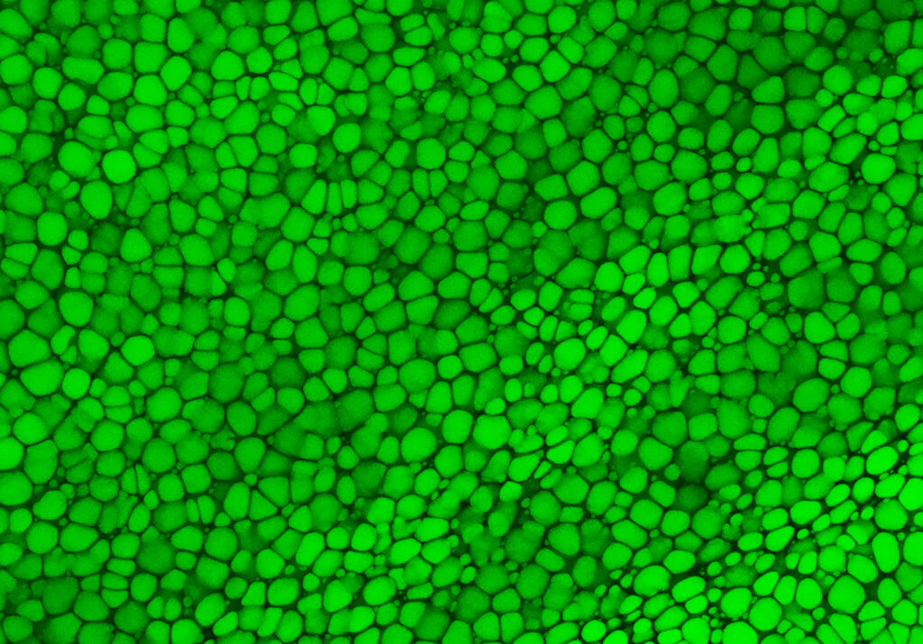
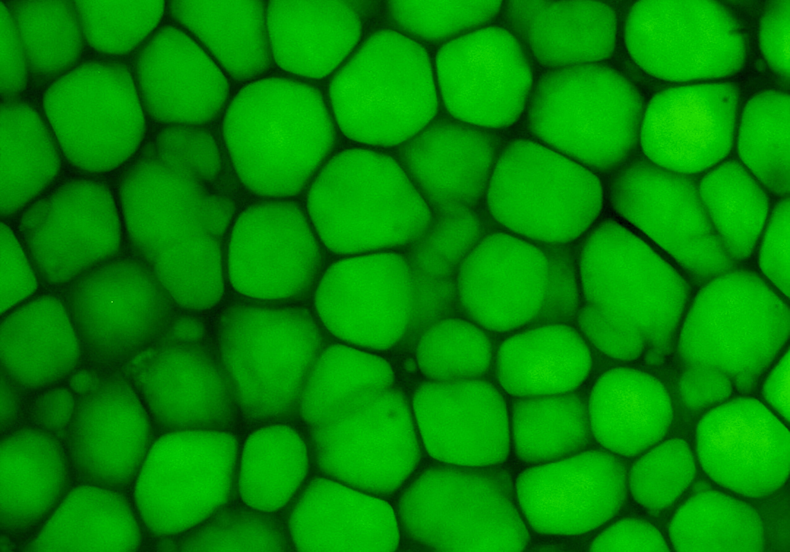
In a study recently published in Science, the team found that LC precursor cells in the ear cartilage of mice start out as being similar to the progenitors of ‘conventional’ cartilage, but then as they undergo terminal differentiation, LC precursors activate lipid metabolism genes, including de novo lipogenesis enzymes that convert glucose to fatty acids. However, unlike fat cells, LCs could not take up additional fatty acids from the circulation, nor break down the stored lipids, resulting in very stable fat-filled bubbles.
Through a large network of collaborators, the investigators identified LCs in the tissues of many phylogenetically diverse mammals, including the intricately shaped ears of the bat, but curiously not in those of non-mammalian species that lack external ears such as amphibians, reptiles, and birds. The team also successfully differentiated human embryonic stem cells into organoids with structures that resembled developing “lipocartilage” found in the face and neck. These findings uncovered a hitherto uncharacterized role of LCs in the development and biomechanics of flexible cartilage, inspiring novel approaches to regenerate cartilage.